For whom and why?
Dr. Gianfermi – Practices in Levallois-Perret and Paris Batignolles
We treat high-risk skin cancers on the face and functional areas using Mohs micrographic surgery (M-O-H-S) or slow Mohs surgery, depending on the indication and type of reconstruction. The entire process is carried out in a short cycle combining dermatological care, plastic surgery, and dedicated histopathology/anatomopathology, with advanced reconstruction on the same day (Mohs) or after margin validation (slow Mohs).
Why this technique?
When every millimeter counts, as is the case for the nose, eyelids, lips, ears, canthus, etc., we must use the most conservative technique for optimal results. The goal is not only to remove the tumor: we must heal while preserving as much healthy tissue as possible. Mohs surgery was designed for this purpose. It allows for exhaustive control of the margins at each stage, guiding re-excision only where the tumor persists, followed by customized reconstruction.
The team and infrastructure
- Center dermatologist: diagnosis, mapping, oncological and skin monitoring.
- Plastic surgeon: Dr. Gianfermi, Mohs/slow Mohs excision and advanced reconstructions (local plasty, flaps, grafts).
- Partner histopathology/anatomopathology laboratory, experienced in Mohs reading (frozen sections) and slow Mohs (paraffin sections with immunomarking).
- Dedicated external operating room, local anesthesia, coordinated care in Levallois-Perret and Paris – Boulevard des Batignolles.
- What is Mohs micrographic surgery?
Mohs (M-O-H-S) is a staged and mapped skin surgery. The visible tumor is removed by taking a very thin margin (usually 1 to 2 mm) around the periphery and depth. The specimen is flattened, stained, oriented, and then immediately analyzed under a microscope. If tumor cells remain anywhere around the edge, we know exactly where and only resect that area. We repeat this process until we achieve healthy margins.
The guiding principle is maximum control of the edge and depth at each stage, with maximum tissue preservation and high cure rates.
What is slow Mohs?
Slow Mohs (delayed Mohs) follows the same principle of complete mapping, but the analysis is performed on paraffin sections—which take longer to prepare but are thinner and more accurate for certain tumors, with the possibility of immunohistochemistry (e.g., SOX-10, MART-1) specifically marking certain tumors. Results are usually available within 24–48 hours. Reconstruction is performed after confirmation of healthy margins. The patient is then left with a temporary dressing while awaiting the results.
For which tumors?
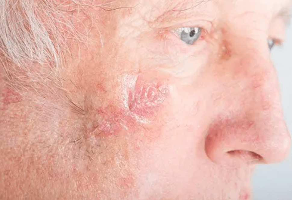
- Basal cell carcinoma (BCC): infiltrating, sclerodermiform, or recurrent nodular forms, poorly defined and located in areas of aesthetic or functional importance (nose, eyelid, ear, lip, etc.).
- High-risk cutaneous squamous cell carcinoma (SCC) (size, high-risk site, differentiation, perineural, recurrence).
- Adnexal tumors (e.g., microcystic adnexal carcinoma) and a few selected rare tumors.
- Protuberant dermatofibrosarcoma (PDFS): benefit of complete margin mapping.
- Lentigo maligna/melanoma in situ of the face: slow Mohs is preferred, with immunomarking.
What is generally not indicated: the Mohs technique is not the gold standard for all skin cancers. For invasive melanoma, standard wide excision according to recommendations remains the rule, with the staged option being discussed on a case-by-case basis. It is also unnecessary if the tumor is in an area where the usual margins do not involve major functional or cosmetic complications.
Why does this technique reduce “safety” margins?
In conventional surgery, standardized margins (ranging from a few millimeters to more than a centimeter, depending on the tumor) are removed from the outset, and then the edges are sampled. With Mohs surgery, we start with 1–2 mm, because the margins are analyzed a few minutes after excision. Further surgery is then performed only where the results are positive. The result is often less tissue loss for at least equivalent oncological control, and often superior control in high-risk tumors.
Mohs surgery procedure (day of surgery)
Marking: photos, marking, consent, local anesthesia.
1st pass:
Tumor + collar of ≈ 1 mm (1–2 mm depending on the site) and thin deep plane.
Mapping & immediate reading: the specimen is flattened, stained, oriented, then passed through the cryostat for reading under the microscope. Typical time frame: 45–90 min.
Result at the chair:
Clear margins: proceed to reconstruction.
Positive margins: identify the location and perform a targeted second pass.
Second pass:
Peripheral removal of the affected area(s), followed by immediate re-examination.
Third pass:
Exceptional.
Reconstruction: continuous (Mohs) — from simple suture to local flap or graft, depending on size and location. Options were discussed preoperatively to align technique and patient priorities.
Usual duration:
1.5 to 3 hours depending on the number of steps and reconstruction.
Mohs cycle
Marking → 1st pass (1–2 mm) → Mapping & reading →
Healthy margins? — yes → Reconstruction
└— no → 2nd targeted pass → Rereading → (rare) 3rd pass
And the slow Mohs procedure
1st pass mapped with narrow margin.
Paraffin + staining (immuno if necessary).
Result on day +1/day +2. If healthy → reconstruction planned quickly. If positive → 2nd targeted pass.
Major advantage: increased accuracy for melanocytic lesions in situ and certain clinically unclear tumors.
Slow Mohs (deferred)
1st pass → Paraffin (+/− immuno) → Results on D+1/D+2 →
Healthy? — yes → Reconstruction
└— no → 2nd targeted pass
Advantages over “conventional” excision
- Complete control of margins at every stage.
- Tissue sparing (less skin is removed), improved function and aesthetics (eyelid, nose wing, vermilion border, helix, etc.).
- Lower recurrence rates in many situations (facial CBC, recurrences, infiltrating forms).
- External, local anesthesia, generally straightforward aftercare.
- Reconstruction integrated into the same care pathway.
Reconstructions: A complete arsenal, a reasoned choice
- We naturally perform cosmetic sutures and local plasty (advancement, rotation, transposition).
- Sometimes flaps (bilobed, nasogenian, fronto-nasal, Mustard, Tenzel, etc.) depending on regional constraints.
- Partial or total skin grafts are reserved for very large losses of tissue. The choice is made on a case-by-case basis, taking into account the size and location of the loss of tissue and the patient’s priorities (discretion, symmetry, maintenance of eyelid or lip function, nasal breathing, etc.).
Tissue-sparing approach
Standard excision: [large oval removed]
Mohs: [smaller final oval] + targeted additions only
Preparing for your visit
Eat lightly; bring something to tide you over between stages.
Bring a list of your medications (anticoagulants/antiplatelet agents).
Arrange for someone to accompany you if more extensive reconstruction is anticipated.
In most cases, you will be able to return home the same day.
Frequently asked questions
1) Is Mohs surgery suitable for all skin cancers?
No. It is primarily indicated for high-risk and/or poorly defined carcinomas. For invasive melanoma, standard wide excision is preferred; staged approaches are discussed in multidisciplinary meetings.
2) Why start with only 1 mm?
Because the reading is exhaustive: we only add where the tumor persists, which spares healthy tissue.
3) How many stages?
Most often one or two. Three is rare.
4) Pain and after-effects?
Local anesthesia. After-effects are generally moderate, requiring standard painkillers and simple local care.
Informative notes
This content is intended for the general public and does not replace a consultation. Indications depend on clinical examination, histology, and location.