Dr. Massimo Gianfermi and Dr Garat use the latest fotofinder technology for screening.
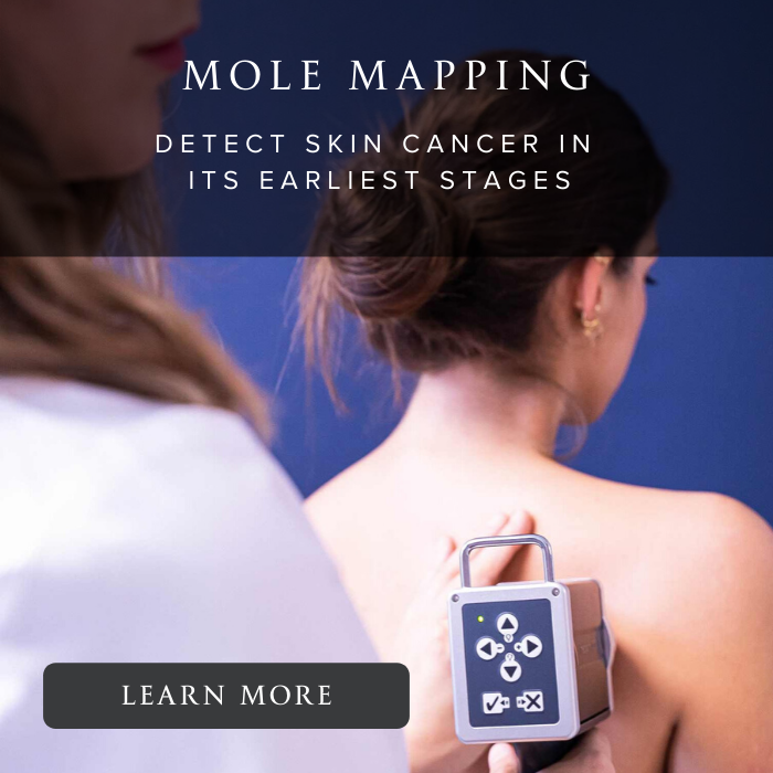
Automated screening with fotoFinder
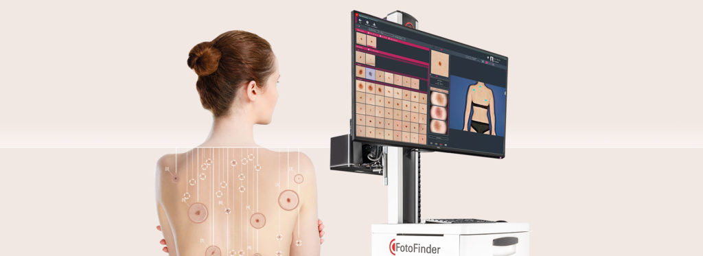
Whole skin diagnosis for early detection of skin cancer
Dr. Gianfermi’s and Dr Garat’s FotoFinder helps you document over time the skin and various melanin spots and recognize, as early as possible, pathological alterations. This can sometimes save lives.
This cutting-edge Total Body Mapping automated technology is based on the “two-step digital evolution control method,” a combination of whole-body photography and dermatoscopy recommended by opinion leaders worldwide for screening at-risk patients.
Excellence & Artificial Intelligence
ATBM - state-of-the-art technology like no other
The ATBM technique is chosen by Dr. Gianfermi and Dr Garat. Based on the “two-step method” for digital tracking, it intelligently combines whole body photography and digital dermoscopy.
ATBM is the only automated polarized light whole body mapping system that documents AND analyzes the entire body surface in minutes. The cross-polarized light whole body photos provide remarkable “macrodermoscopic” information on the structure of individual lesions.
The unique “Bodyscan” helps you detect new and evolved nevi quickly and automatically. The Moleanalyzer software gives a malignancy score on each individual nevus.
Request an appointment
Submit your appointment request and one of our patient care coordinators will contact you shortly.
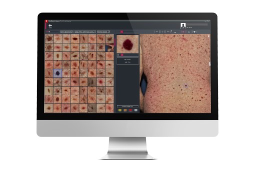
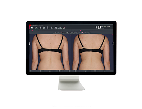
Bodyscan master with Mosaic View
The Bodyscanmaster identifies new and altered skin lesions, extracts melanin spot icons in the overall body photo, and ranks them according to relevance
True video dermatoscopy with CrystalView technology
Full HD video camera medicam 1000s seamlessly integrated with live optical zoom and variable magnification
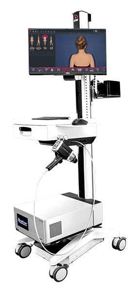
Really mobile ATBM tower
Turnkey ATBM tower installed, truly mobile, with high quality, high performance FotoFinder hardware.
Artificial Intelligence (AI)
The AI score of the Moleanalyzer pro supports analysis and risk assessment of skin lesions. ATBM master further integrates this technology to optimize total body dermatoscopy results.
Very high resolution images with PolFlash XE
The ATBM master procedure provides very high resolution, polarized, RAW-processed images. The PolFlash XE computer-controlled xenon flash enables both cross-polarized medical imaging and studio lighting for aesthetics.
Ultra-fast and delegable imaging procedure
The ATBM master is equipped with the best processing unit we have ever built. The process is extremely fast, intuitive and delegable.
Fotofinder the reference for global imaging
Artificial intelligence for lesion assessment
Dr. Gianfermi and Dr Garat are using the new ATBM master that makes whole body dermatoscopy possible for the first time. The combination of the “whole skin” imaging procedure with artificial intelligence and new software features for lesion visualization will improve diagnostic accuracy.
FotoFinder Bodyscan
New and changed lesions detected at a glance
Dr. Gianfermi’s and Dr Garat Bodyscan ATBM has redefined the comparison of before and after photos. The expert system, integrated and fully automatic, detects new lesions and those that have evolved, as soon as the photos are taken. The results are immediately available, and the diagnosis is simply more reliable.
About
Get aDiagnosis
Early detection of skin cancers offers better chances of cure, especially in the case of cutaneous melanomas.
In France, there is no organized screening program for skin cancers. Their early detection therefore relies either on your doctor’s initiative, or on yours if you have spotted a potentially suspicious lesion (wound that does not heal, pimple or scab that persists or evolves, brown “spot”), or a mole “different from the others”.
On average, 100,000 skin cancers are diagnosed each year.
Testimony of Dr. Gianfermi
About FotoFinder, Interview conducted in Paris, France
I think the ATBM bodystudio from FotoFinder has the potential to change the way we do digital monitoring. Typically, we select lesions with a manual dermatoscope and then image and monitor them. The potential I see in this machine is that instead of focusing on a single lesion,I look at the whole body and then do the side-by-side comparison because I don’t need to go further into the morphology of an individual globule.
I need to know if a lesion is evolving symmetrically or asymmetrically and that’s the information I get with whole-body photography in polarized light.